Site-Rite™ 8 Ultrasound System

site-rite_8_hero.png
Loading
Overview

Simplifying Vascular Access

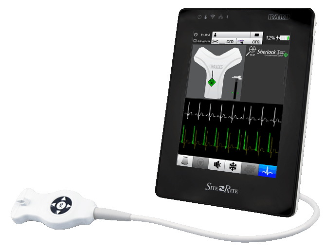
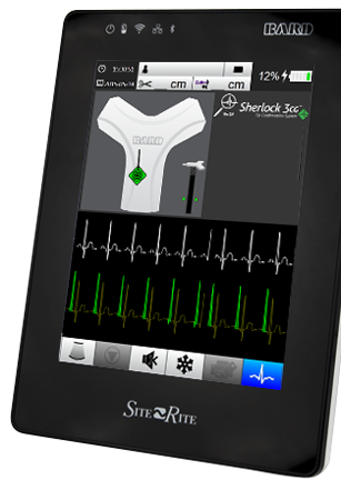
The Site-Rite™ 8 Ultrasound System with integrated SHERLOCK 3CG™ Diamond Tip Confirmation System (TCS) is designed to be simple and intuitive. Our ultrasound and imaging technologies help you visualize vessels, needle trajectory, and final tip location.

true
true
true
true
Features and Benefits
Making the Invisible Visible

The Site~Rite™ 8 Ultrasound System with integrated SHERLOCK 3CG™ Diamond TCS is designed to simplify vascular access by allowing you to visualize PICC placements from beginning to end.

Traditional ultrasound imaging systems allow you to:

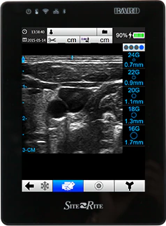
Visualize the vessel and surrounding anatomy

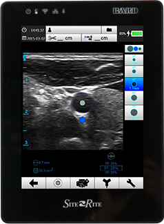
Visualize and measure catheter size relative to the vessel
Visualize virtual catheter location during PICC placement

However, the Site~Rite® 8 Ultrasound System with Pinpoint™ GT Needle Technology and integrated SHERLOCK 3CG™ Diamond TCS uses traditional ultrasound imaging and a portfolio of visualization products, enabling you to:

Visualize the needle trajectory

Visualize maximum P-wave to confirm final catheter tip location

Getting to the Heart of the Matter

Using X-ray to confirm PICC tip placement wastes clinical care time, exposes patients to harmful radiation, and delays medical therapy. The SHERLOCK 3CG™ Diamond Tip Confirmation System (TCS) gets to the heart of the matter by using an adult patient’s cardiac electrical activity to position the tip of the PICC in close proximity to the cavoatrial junction

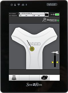
Visual catheter tip tracking

Real-time catheter tip tracking technology is used to display both catheter direction and orientation. This helps to increase placement efficiency and reduce catheter malpositions as compared to “blind” catheter placements.

Visual catheter tip confirmation

The SHERLOCK 3CG™ Diamond Tip Confirmation System (TCS) is an advanced technology that deciphers P-wave morphology and determines PICC tip position relative to the CAJ.

Simultaneous views of both catheter tip tracking and the patient’s electrocardiogram (ECG) are displayed, allowing you to navigate the catheter tip into the SVC and confirm catheter placement in proximity to the CAJ using ECG technology.

Designed to minimize the challenges of the PICC placement process by helping:

  • Locate and navigate the catheter tip in real-time
  • Confirm catheter tip placement at the CAJ in adult patients with identifiable P-waves
  • Eliminate confirmatory chest X-rays to reduce radiation exposure
  • Release the line to provide life sustaining therapy at the bedside
  • Reduce time wasted waiting for PICC tip confirmation using alternative post-placement methods

Real-Time Needle Visibility

  • Pinpoint™ GT Needle Technology on the Site~Rite™ 8 Ultrasound System is designed to improve the ultrasound-guided needle insertion experience by providing visual feedback of needle trajectory and needle tip positioning in relation to the structure of interest.

The Pinpoint™ GT Needle Technology allows you to:

  • Visually align the needle pathway, prior to insertion
  • Visualize needle tip location in relation to the target structure
  • Confirm needle intersection of the ultrasound plane
  • Maintain image integrity by eliminating the need to move the probe to find the needle
  • Place needles out-of-plane, in-plane, or oblique to the target structure

How it works

Making Virtual Needle Visibility a Reality

The Pinpoint™ GT Safety Introducer Needle

  • The Pinpoint™ GT Safety Introducer Needle emits a passive magnetic field

Sensors within the Pinpoint™ GT Linear Ultrasound Probe

  • Sensors within the Pinpoint™ GT Linear Ultrasound Probe detect the location and position of the needle’s magnetic field

Pinpoint™ GT Software on the ultrasound system

  • Pinpoint™ GT Software on the ultrasound system interprets the data to create a virtual needle image on the ultrasound display
Pinpoint GT Safety Introducer Needle Product Codes

Description

L x WTaperThicknessProduct CodeQty/Box
48 inch Probe Cover Kit with 21G Pinpoint™ GT Safety Introducer Needle48” x 6”None1.0 mil97706225
Pinpoint™ GT Safety Introducer NeedleNANANA97706245

Warning: Pinpoint™ GT Needle Technology is not qualified for use with Site~Rite™ Needle Guides. Using Site~Rite™ Needle Guides with the Pinpoint™ GT Needle Technology may result in injury to the patient.

Products & Accessories
RELATED PRODUCTS NOT AVAILABLE

Medical Equipment Roll Stand (MER)

Designed to simplify medical equipment transportation.

The mounting plate & equipment mount supports medical equipment up to 15 lbs, including qualified Site~Rite™ Ultrasound Systems.

Please see your Ultrasound System Instructions for Use for the appropriate rollstand qualified for use with your device.

Articulating equipment mount supports medical equipment up to 8 lbs, including SHERLOCK 3CG™ TCS.

Product codes:
9770116 - Medical Equipment Roll Stand (MER)
9770109 - Laptop mounting tray (for laptop medical equipment)
9770133 - SHERLOCK 3CG™ TCS for the MER
MW260MFIC129 - Brother™ printer for the SHERLOCK 3CG™ TCS system
Resources

Needle Guide Kits

BD offers an exclusive line of needle guides designed for Site-Rite™ Ultrasound Systems to help clinicians guide needles to vascular structures at specific depths.

Pinpoint™ GT 21G Needle Guide Kit Features and Benefits

Product Codes

Description

L x W

TaperThicknessDepths (cm)0.51.01.52.02.53.03.5Product
Code
Qty
Per
Box
21G Needle Guide Kit48” x 6”None1 mil   97706905
21G Needle Guide Kit96” x 6”6” to 5.5”1 mil   97706915
21G Needle Guide Kit48” x 6”None1 mil    97706925
21G Needle Guide Kit96” x 6”6” to 5.5”1 mil    97706935
  • Funnel design to aid in needle insertion
  • 0.5 – 3.5cm depth coverage
  • Guides needle to specified depth under probe
  • Available with 48" and 96" probe covers

Site~Rite® Needle Guide Kit Features and Benefits

Pinpoint GT Safety Introducer Needle Product Codes

DescriptionL x WTaperThicknessDepths (cm)0.51.01.52.02.53.03.5Product
Code
Qty
Per
Box
21G Needle Guide Kit48” x 6”None1 mil    900013B0110
21G Needle Guide Kit96” x 6”6” to 3”1.5 mil    900219610
20G Needle Guide Kit48” x 6”None1 mil   9001C021410
20G Needle Guide Kit96” x 6”6” to 3”1.5 mil   900209610
18G Needle Guide Kit48” x 6”None1 mil    9001C021210
18G Needle Guide Kit96” x 6”6” to 3”1.5 mil    900189610
  • Guides needle to specified depth under probev
  • Available in 18G, 20G, and 21G
  • Available with 48" and 96" probe covers

Warning: Pinpoint™ GT Needle Technology is not qualified for use with Site~Rite® Needle Guides. Using Site~Rite® Needle Guides with the Pinpoint™ GT Needle Technology may result in injury to the patient.

Probe Cover Kits

BD offers a complete line of sterile probe cover kits for Site-Rite™ Ultrasound System probes.

Sterile ultrasound probe covers help reduce the potential for cross-contamination and bloodstream infections by creating a sterile barrier between the ultrasound probe and the patient.

Features and Benefits

  • Designed for easy application
  • Available in 12, 48 and 96-inch lengths

Pinpoint GT Safety Introducer Needle Product Codes

DescriptionL x WTaperThicknessProduct CodeQty/Box
12 inch Probe Cover Kit12” x 5”None2.0 mil900001210
48 inch Probe Cover Kit48” x 6”None1.0 mil9001C019710
96 inch Probe Cover Kit96” x 6”6” to 3”1.5 mil900009610
Intraoperative Probe Cover Kit12” x 5”None.0 mil900023B0110
96” x 6”6” to 3”1.5 mil  
Reference

To learn more about how the Site~Rite™ 8 Ultrasound System with Pinpoint™ GT Needle Technology and integrated Sherlock 3CG™ Diamond TCS can be an asset in your facility, click the “Contact Us” button near the top of the page to request more information from a clinical representative in your area.

true

BD-20006

true
Site-Rite™ 8 Ultrasound System BD Site-Rite™ 8 초음파 시스템 Making the Invisible Visible,

The Site~Rite™ 8 Ultrasound System with integrated SHERLOCK 3CG™ Diamond TCS is designed to simplify vascular access by allowing you to visualize PICC placements from beginning to end.

,Traditional ultrasound imaging systems allow you to:,

Visualize the vessel and surrounding anatomy

,Visualize and measure catheter size relative to the vessel,,Visualize virtual catheter location during PICC placement,,,

However, the Site~Rite® 8 Ultrasound System with Pinpoint™ GT Needle Technology and integrated SHERLOCK 3CG™ Diamond TCS uses traditional ultrasound imaging and a portfolio of visualization products, enabling you to:

/content/dam/bd-assets/bd-com/en-us/logos/bd/header-bd-logo.svg